Antibodies vs minibinders
While framework-free minibinders, often designed through structural hallucination, represent a truly groundbreaking advance in bioengineering and offer solutions where conventional binders fall short, in therapeutic development the antibody scaffold still carries advantages that are hard to reproduce from scratch. Its structure has been characterized in exhaustive detail. Its developability liabilities, such as aggregation-prone patches, deamidation/isomerization hotspots and oxidation-sensitive residues, are catalogued and screened for routinely. It has a well-understood immunogenicity profile, a naturally long serum half-life that can be extended further by engineering FcRn engagement, and a deep bench of validated bispecific formats – just to name a few.
In other words, the antibody framework is here to stay, and the cloud lab service to turn antibody sequences to functional data must be optimized for it.
Mammalian vs cell-free expression
Antibodies are notoriously hard to fold in E. coli-based expression systems, even as single-chain Fv fragments, primarily due to the large number of disulfide bonds that must be formed in correct pairs. In contrast, CHO- and 293-based cell lines have been the industry workhorse for decades to produce antibodies and antibody fragments. The mammalian ER is an oxidizing compartment stocked with protein disulfide isomerases that both form and reshuffle disulfide bonds, with BiP and the calnexin/calreticulin system to hold folding intermediates, and with peptidyl-prolyl isomerases to catalyze the slow proline isomerization. More importantly, it runs an assembly checkpoint. The CH1 domain of the heavy chain is intrinsically disordered on its own and only acquires its immunoglobulin fold on pairing with a folded light-chain constant (CL) domain; until that happens, BiP binds it and the chain is retained[1]. Secretion is therefore conditional on correct assembly.
The idea of using cell-free protein synthesis (CFPS) to produce antibody-like binders such as VHH and scFv has regained momentum in recent years in the context of rapid testing of binder function following computational sequence design. The appeal is understandable. On the gene-synthesis side, clonal plasmids are not necessary for CFPS and much cheaper and faster gene fragments are sufficient. On the protein-expression side, a few hours of incubation appears to produce a sufficient amount of binder protein for binding assays on SPR or BLI. On the binding-assay side, the binder protein produced in CFPS, with or without purification, can be loaded onto the solid surface via an affinity tag fused to the scFv, ready for the target protein to bind.
Given the challenge of scFv expression in E. coli-based CFPS systems, various additives such as chaperones and artificial redox systems have been added to CFPS systems. Nevertheless, the fraction of well-folded protein remains poorly characterized in most commercial CFPS systems, let alone predicted accurately for a new binder sequence.
The risks of sub-optimal folding
The risk of sub-optimal folding of the antibody fragment manifests in different ways in BLI/SPR experiments depending on whether the binder or the target is immobilized on the sensor surface. Let’s unpack this.
Immobilizing target, binder in solution
It is common in conventional BLI and SPR protocols to see that the target protein is immobilized, and the binder is used in solution. In this orientation, we have to know the concentration of the well-folded binder to calculate $k_\text{on}$: the association phase approaches its plateau at an observed rate $k_\text{obs} = k_\text{on}[\text{Binder}] + k_\text{off}$, so that $k_\text{on} = (k_\text{obs} - k_\text{off})/[\text{Binder}]$, with $[\text{Binder}]$ the well-folded binder concentration. I stress ‘well-folded’ because not all binder protein is well-folded. Common methods based on UV-absorption, dye staining and tag quantification cannot tell whether the binder is well folded. If 10% of the binder is well folded, then $[\text{Binder}]$ is only 10% as much as you think it is, and $k_\text{on}$ is 10-fold higher than you thought.
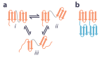
There are many ways for antibodies and engineered fragments (e.g., scFv) to misfold. Even when each of the variable domains is itself folded, they may not come into contact in the natural orientation (Fig. 1a, ii vs i). Of course each variable domain can misfold on its own (Fig. 1a, iii vs i). The open state (ii) is the entry point to the two classic scFv failure modes: aggregation, and inter-chain pairing in which the VH of one molecule pairs with the VL of another to give diabodies and higher multimers, a tendency set largely by linker length[2]. The multimers are the more insidious of the two in a binding assay, because they are soluble, they bind, and they are multivalent.
Another practical problem is that high concentration of binder is needed. E.g., in initial screening, the $K_\text{D}$ may be >100 nM, and during the association, the binder concentration must be at least 2 to 3 times the $K_\text{D}$ to see the association curve plateau over time to fit the $k_\text{obs}$ parameter. This means you need the binder at a concentration of at least ~500 nM. The expression system may not have a high enough yield to reach that total binder concentration, let alone that well-folded fraction.
Immobilizing binder, target in solution
What if we flip the orientation, i.e., having the binder immobilized on the probe, and the target in solution? On the surface this appears to be a great idea. First, the amount of binder protein needed is relatively small, 20 - 50 nM may be more than enough. Second, we don’t need to know the concentration of the correctly folded binder protein. Of course, this orientation requires knowing the concentration of the well-fold target protein. Fortunately, since the same lot of the target protein is used to evaluate multiple binders, the well-folded proportion is the same across these binders, so at least the relative $k_\text{on}$ values across the binders tested are still trustworthy. In addition, target proteins are usually produced in large batches and undergo extensive QC at providers, so the well-folded proportion probably falls in a narrower range than for the binders, especially if a hard-to-fold binder is expressed in CFPS.
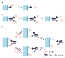
So what’s the downside? The danger arises when the target protein exists in a dimer or oligomer format, which is common. In addition to target proteins that are naturally dimeric or oligomeric, many target proteins, especially secreted proteins and ectodomains of human transmembrane proteins, are produced as Fc fusions. The Fc domain is a dimer, leading to the presence of two copies of the target protein in one molecule. When this happens, the target protein may engage with two copies of the binder protein that are immobilized on the SPR/BLI surface, leading to inflated affinity (see this post for detailed analysis, but its figure is reproduced as Fig. 2 for readers’ convenience).
The best practice
Considering all the pros and cons above, we propose the following guideline.
When the target protein is naturally monomeric and is available in non-dimerizing form (e.g., untagged or His tagged, as opposed to Fc-tagged), we should immobilize the binders and take advantage of the small binder amount requirement and the controlled and uniform quality of the target protein.
However, when the target protein is dimeric/oligomeric either naturally or artificially (e.g., as Fc fusion), we recommend immobilizing the target and produce monomeric binders with the production method that offers the highest folding quality. This means we should use CHO or 293T cells for secretory expression. For format, we recommend using the Fab format over scFv or full length antibody. Compared to scFv, where the VH and VL are held by a simple flexible linker and rely on the natural affinity between VH and VL to align the two domains properly, in Fab the constant domains (CH1 on the heavy chain and CL on the light chain) dimerize with an inter-chain disulfide bond, facilitating the alignment of VH and VL (Fig. 1b). Direct comparisons show that the Fab format can rescue variable domains whose scFv counterparts are poorly behaved[3]. Compared to a full-length antibody, the Fab is monomeric, avoiding the bivalent binding complication discussed above.
Maximizing speed and quality at affordable cost
But what about the cost and time of the CHO/293T system? In the past, making the plasmid (about 12 to 15 US cents per base) is indeed about twice as expensive as gene fragments (about 5 to 7 US cents per base). And expression of antibody in the CHO/293T cells takes 5 to 10 days to reach maximum yield. Luckily, RootPath has honed its knife on gene synthesis for many years and has lowered the cost of plasmid synthesis to be in line with gene fragments from most providers (about 5 to 7 US cents per base, with the rapid synthesis workflow for rapid testing; high-throughput synthesis offers even lower price). We have also optimized cell lines and growth conditions of CHO/293T cells so that a sufficient amount of Fab can be produced in as little as 1.5 days. When Fab is used in solution, which requires its concentration to be known, we use a well-optimized method to purify the Fab for accurate total concentration measurement.
Taken together, you can test CHO/293T-produced, purified, purity-tested Fab at $150 to $200 per sequence depending on batch size, with a turnaround time as short as 6 days. If you are designing antibody-like sequences, be sure to check out the Fab pipeline on BinderVerse.
As always, feel free to email me at [email protected] or book an online meeting to discuss your projects.
– Xi Chen
References
- Feige MJ, et al. (2009). An Unfolded CH1 Domain Controls the Assembly and Secretion of IgG Antibodies. Molecular Cell 34(5):569-579. doi:10.1016/j.molcel.2009.04.028
- Holliger P, et al. (1993). "Diabodies": small bivalent and bispecific antibody fragments.. Proc. Natl. Acad. Sci. U.S.A. 90(14):6444-6448. doi:10.1073/pnas.90.14.6444
- Röthlisberger D, et al. (2005). Domain Interactions in the Fab Fragment: A Comparative Evaluation of the Single-chain Fv and Fab Format Engineered with Variable Domains of Different Stability. Journal of Molecular Biology 347(4):773-789. doi:10.1016/j.jmb.2005.01.053